Methods of Investigation
for FP7 partners
and coordinators
|
|||
| Home | |||
| Cytotechnology Group | |||
| Staff | |||
| Methods | |||
| Projects | |||
| Research Results | |||
| Patents | |||
| Image Gallery | |||
| Cooperation | |||
| Contact Us | |||
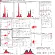
Viability Test Using AnnexinV-FITC and PI |
||||||||||||||||||||||||||||||||||||||||||||||||
|
||||||||||||||||||||||||||||||||||||||||||||||||
| COOPERATION NEWS | ||
FP7 Programme involves subjects
|
||
| CURRENT RESEARCH | ||
|
||
| 2005-2007 © LBMI |


